Mastering Head & Neck Anatomy: 2d Radiographs And 3d Images
Published 2/2026
MP4 | Video: h264, 1920x1080 | Audio: AAC, 44.1 KHz, 2 Ch
Language: English | Duration: 3h 48m | Size: 7.21 GB
A Complete Guide to Learn Head, Maxillofacial and Neck Anatomy: Focused on Dentistry but Also Useful to Medical Students
What you'll learn
Locate All Head and Neck Anatomical Landmarks, Including bone, muscles, nerves and blood vessels on representative images of the body, such as in Anatomy Books.
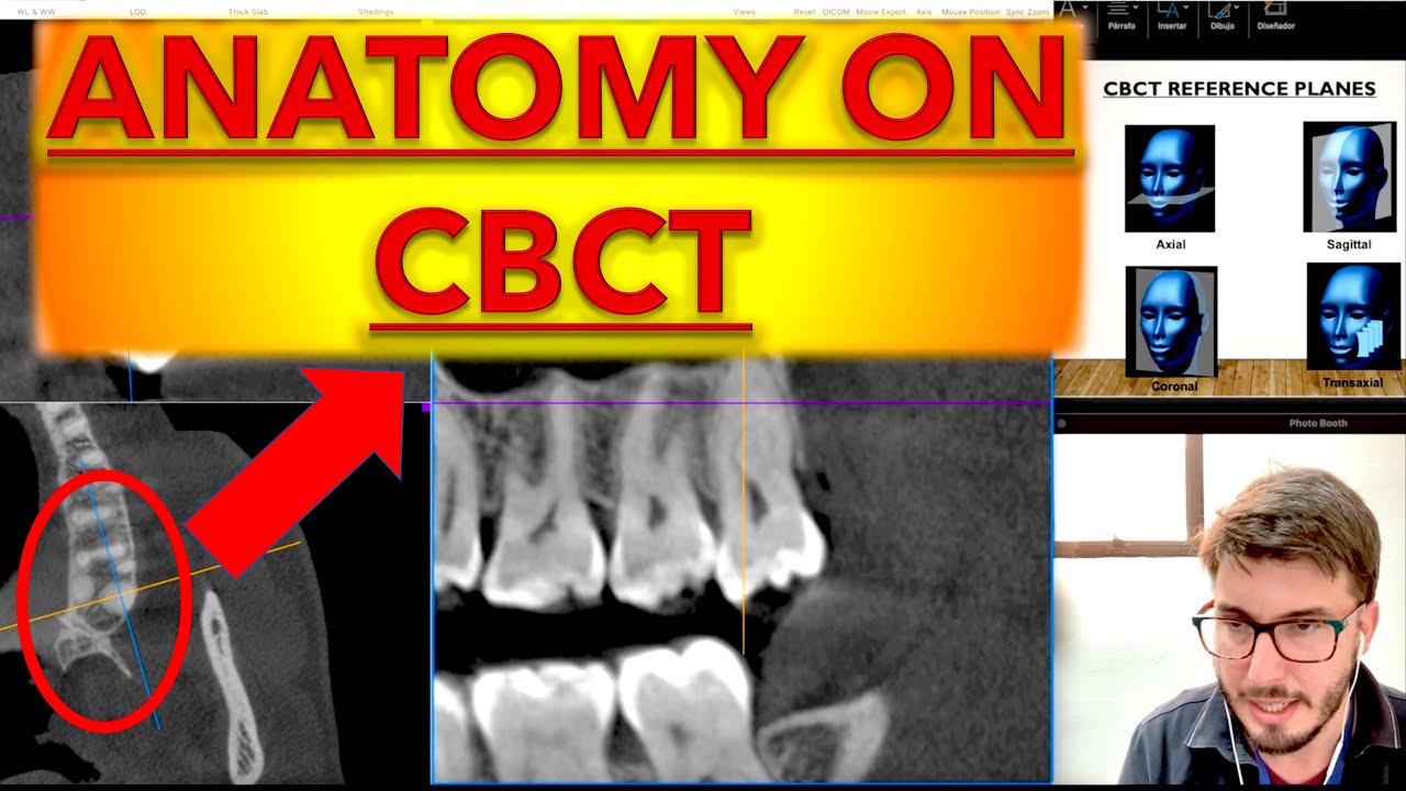
Understand How to Find these Anatomical Landmarks in 2D Intraoral and extraoral radiographs
Find all anatomical structures in 3D on medical CT, CBCT, and MRI, correlating with their appearances on 2D planes
Correlate the location of the anatomical structures with their most common clinically relevant alterations, mostly in dentistry, but also for head and neck.
Requirements
Be a Dental or Medical Student
Be a Dentist.
Description
This course provides an intensive exploration of the complex architecture of the human head and neck. By bridging the gap between gross anatomy and modern diagnostic imaging, students will develop a "3D mental map" essential for clinical practice. We move beyond traditional textbooks to examine how physical structures translate into the radiographic shadows of 2D X-rays and the volumetric data of CBCT and CT scans.
What You Will Learn
• Structural Foundations: Detailed study of the osteology, myology, and main blood vessels, as well as neurovascular pathways of the head and neck, with a special focus in dentistry.
• The Art of Interpretation: Identifying anatomical landmarks on 2D projections (panoramic and cephalometric radiographs) and understanding the effects of superimposition.
• Volumetric Analysis: Navigating 3D imaging (CT/CBCT/MRI) across axial, sagittal, coronal and 3D planes to locate anatomical variations and alterations.
• Clinical Correlation: Applying anatomical knowledge to real-world scenarios, including surgical planning, local anesthesia landmarks, and dental surgery.
1. Identify major and minor anatomical landmarks of the head and neck, as well as dental anatomical landmarks with accuracy.
2. Differentiate between normal anatomical variants and potential alterations/pathology on 2D radiographs.
3. Navigate a 3D software environment to isolate specific structures like the mandibular canal or paranasal sinuses.
4. Synthesize 2D and 3D data to create a comprehensive anatomical assessment for clinical use.
Who this course is for
Mostly for Dental Students and Dentists. Also Useful for Medical Students.